Sample preparation instructions
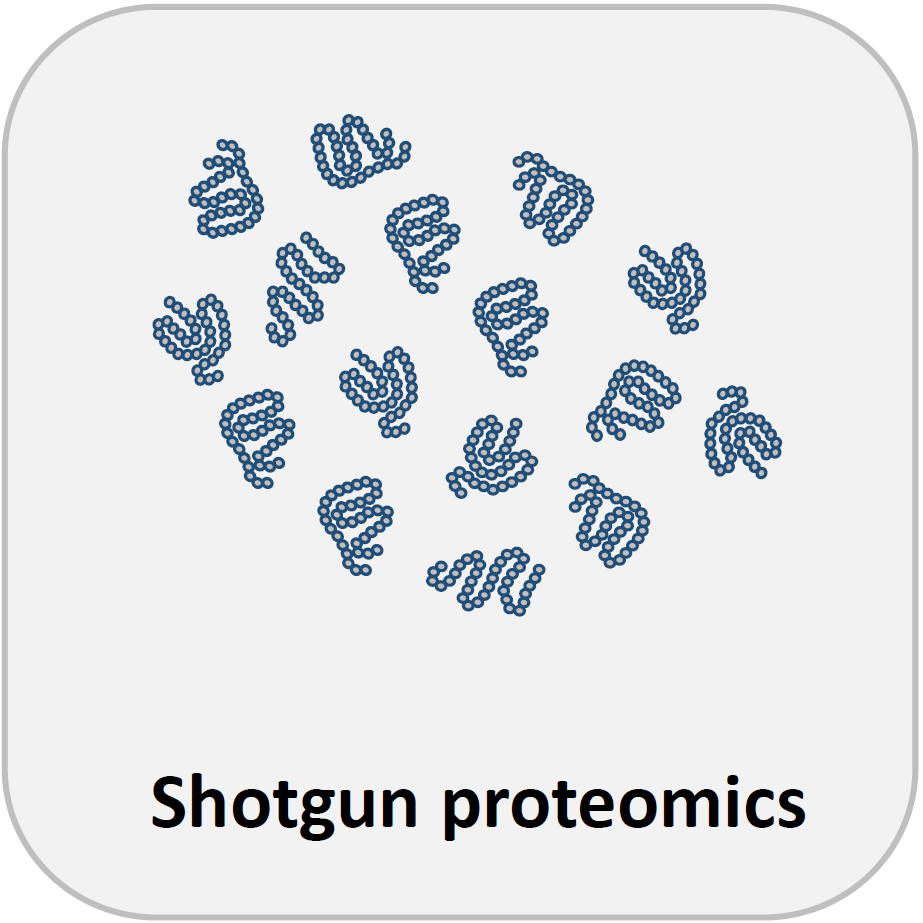
1. Shotgun proteomics
Please provide frozen cell pellets or dissected tissue material, washed with PBS before freezing to remove media or blood components.
For quantitative analysis, at least three replicate samples should be provided for each condition. Cell pellets or tissue samples should contain minimum 30,000 cells, or an amount sufficient to extract ~3 µg of protein material. If cell numbers are not limiting, 1 to 5 million cells/pellet is preferred.
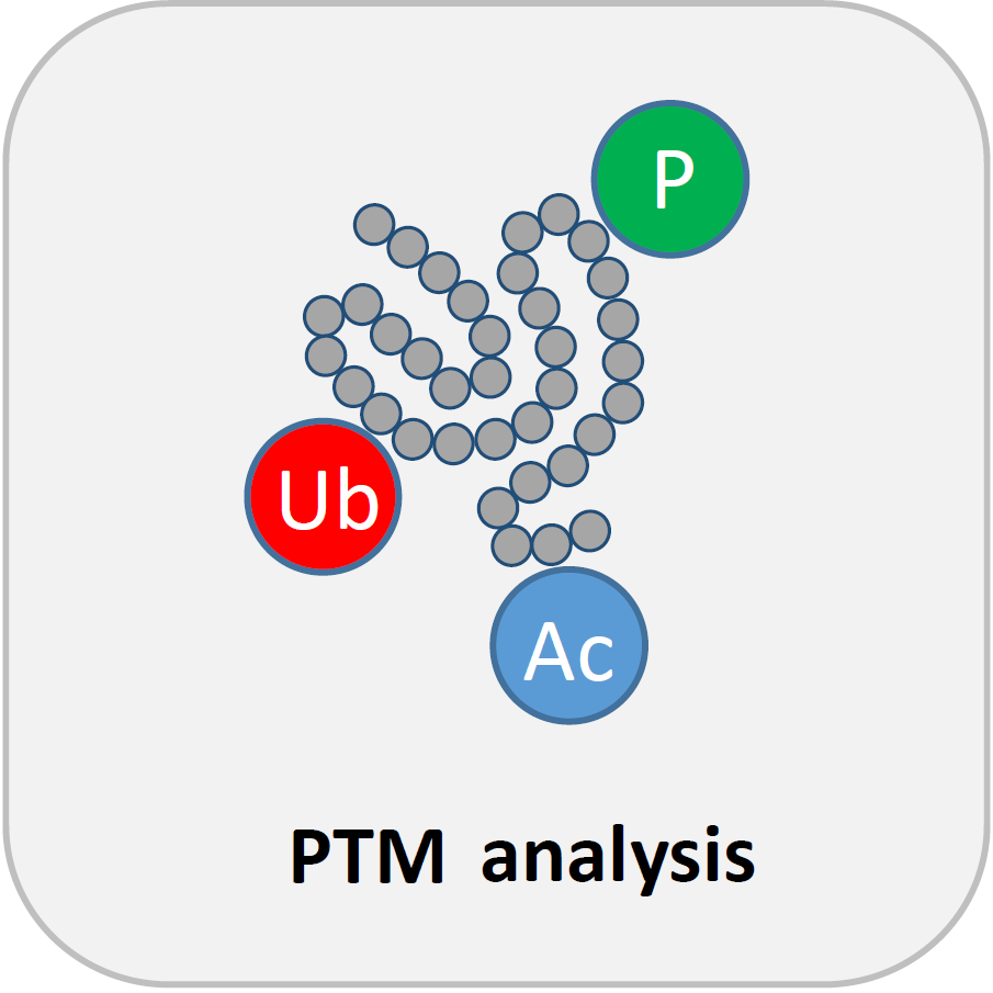
2. PTM analysis
Same instructions apply as for shotgun proteomics, but more input material is required. Please provide frozen cell pellets or dissected tissue material sufficient to extract minimum 2 mg of protein for phosphoproteomics and 10 mg of protein to identify ubiquitin and lysine acetylation sites.
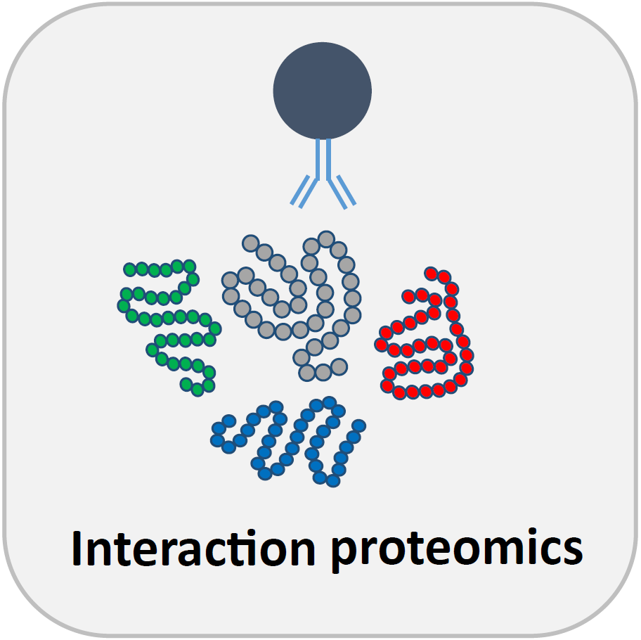
3. Affinity Purification MS (AP-MS)
In most cases you can use the AP protocol of choice, as long as you wash the beads three times in 1 ml trypsin digestion buffer (20 mM Tris.HCl pH 8.0, 2 mM CaCl2) before sending them to us.
For streptavidin beads, either:
o After washing of the beads (according to your protocol) you can incubate the washed beads with maximum 50 µl of 5% SDS in 50 mM TEAB pH 8.5 for 10 minutes at room temperature, facilitating elution by pipetting up-and down the beads regularly. After elution, freeze the eluate and send to us on dry ice
For any other beads, either:
o After the last wash step, re-suspend the beads in maximum 150 µl trypsin digestion buffer, freeze and deliver on dry ice
Comment:
For PTM detection, make sure you add appropriate inhibitors during lysis and IP (not during the washes!), for instance phosSTOP.
For label-free analysis, at least three replicate samples should be provided for each condition, e.g. IP from WT cells vs. IP from KO cells. A negative control condition (no pull down or absence of the bait molecule) is always required to distinguish specific from unspecific binders by labelfree quantitative analysis. Replicate samples are often generated after lysis, by splitting a single lysate in three aliquots for IP. True biological replicates starting from three different cell lysates/culture dishes are also possible.
For each IP replicate sample, we recommend to start from a 2 ml lysate at a concentration of 1.5 mg/ml total protein (3 mg total protein). These values are only an indication and are far from absolute (optimal conditions vary for each experiment), they just give an indication about the amount of material that is typically use for these kind of experiments.
Finally, it is recommended to keep the amount of beads per replicate samples as low as possible, since the antibody/streptavidin/protein A/G will also be cleaved by trypsin leading to additional peptides in the samples. Use maximum 20 µl of settled beads for each replicate if possible.
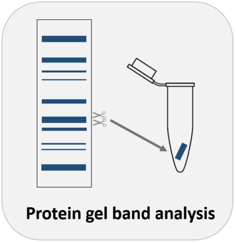
4. Protein Gel Band Analysis
You can run the gel using the system that you commonly use in the lab. For optimal separation, it might be worth to consider a gradient gel (e.g. 4-12%).
Also, commercial pre-cast gels are generally recommended to avoid keratin contamination.
After running the gel, rince the gel with MQ water, stain with colloid coomassie (e.g. simply blue from , Thermo) and take a picture of the gel.
Then, cut out the gel bands using a clean scalpel (under the laminar flow to avoid keratin contamination) and indicate cuts on the picture.
Transfer the gel pieces (with a clean pincet) to individual Eppendorf tubes and rince the pieces once with MQ water (it is easiest to pre-fill the tubes with 500 µl of MQ water). Remove all the water and freeze the gel pieces at -20°C. Send to us on dry ice.